3D printing has opened doors in many fields ranging from medicine, aerospace, construction and cultural conservation. Now, researchers at the University of Minnesota have found a way to use 3D printing for repairing spinal cord injuries.
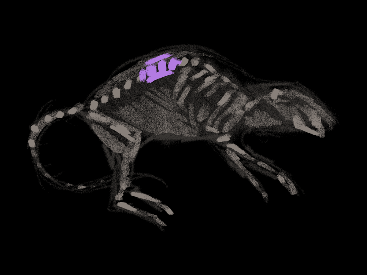
In recent experiments, the researchers used 3D-printed scaffolds with stem cells to help the rats regain function after their spinal cord severing. This development may one day translate into treatment for people living with paralysis.
The research used tiny, lab-grown organs known as organoids to create a 3D printed scaffold. It functions as a biological bridge placed at the site of the spinal cord injury.
The scaffolds were developed with microscopic channels that guide the growth of specialized stem cells known as spinal neural progenitor cells. These NPCs come from human adult stem cells that can divide and mature into nerve cells.
“We use the 3D printed channels of the scaffold to direct the growth of the stem cells, which ensures the new nerve fibers grow in the desired way,” Guebum Han, a former postdoctoral mechanical engineering researcher at the University of Minnesota coauthor of the study, said. “This method creates a relay system that when placed in the spinal cord bypasses the damaged area.”
The scaffolds support cell growth and integration once implanted into rats with severed spinal cords. Within 12 weeks, the stem cells matured into neurons and extended nerve fibers both toward the head and toward the tail.
The new tissue then forms organized bundles of axons, the long and thread-like parts of neurons that transmit signals. It is important to note that the cells not only survived long term, but also developed functional connections with the rats’ own spinal circuits.
The implants allowed for the regrowth of spinal tissue across the injury site by reconnecting the two severed ends of the cord. The boost in movement of the rats’ hind legs suggests that the regenerated nerve fibers were transmitting signals. The blood vessels also established a key circulatory network to sustain the new tissue by growing into the scaffolds.
This discovery represents a major step forward for people with spinal cord injuries, of which there are more than 300,000 people in the U.S. alone.
Until now, one of the greatest challenges has been that the nerve cells die after injury and fail to regrow across the injury site. The Minnesota researchers showed a promising new way to reverse paralysis by creating the scaffold structure with stem cells rather than managing its effects.
However, there are many hurdles to overcome before this method can be tested on humans. Chronic spinal cord injuries differ from recent ones and the scaffold might need to be adapted to work in long-term cases.
Next, researchers will likely need to use patient-specific induced pluripotent stem cells to avoid immune rejection. The polymers used for printing need to be improved to better mimic the spinal cord environment.
Also, additional proteins might need to be incorporated to support long-term axon growth and cell survival. Ensuring a sufficient blood supply to the implanted tissue is another factor.
Overall, there are many challenges that face the transition of the 3D scaffold to human applications, but the study marks a major milestone in regenerative medicine.
It goes to show how engineering and biology can combine to tackle medicine’s most daunting challenges and offers hope that one day people with spinal cord injuries can regain movement and independence.
Categories:
Reversing paralysis using 3D printing
September 15, 2025
More to Discover


