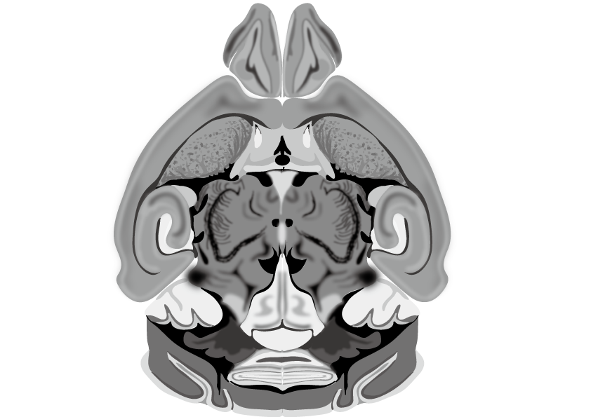
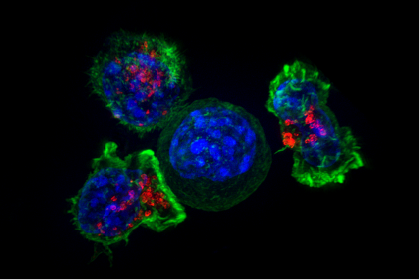
In a landmark study published in Nature, researchers from the Allen Institute at the Baylor College of Medicine and Princeton University achieved a feat once deemed impossible: mapping every single synaptic connection and recording the activity of over 84,000 neurons, all within a cubic millimeter of mouse brain tissue.
It may seem unfathomable, but a sliver of cerebral cortex no larger than a grain of sand now serves as the most detailed wiring diagram of a mammalian brain ever constructed.
The dataset, known as MICrONS, not only visualizes a staggering 500 million synapses across 200,000 cells, but also links those connections to real-time neural activity while mice watched videos of movies like The Matrix and Mad Max: Fury Road.
To build this unprecedented 3D map, researchers started by recording how neurons in the visual cortex responded to visual stimuli.
Then, they carefully sliced the same chunk of brain tissue into nearly 28,000 ultra-thin sections and imaged each slice with electron microscopes over six months.
Finally, using machine learning, they stitched those images together into a digital reconstruction of axons, dendrites and synapses, allowing scientists to “walk through” the cortical wiring as if they were using a GPS system.
“It really has been one of the holy grails of the field from the beginning,” Clay Reid, a senior investigator at the Allen Institute, said in an interview with GeekWire. “There are many thousands of neuroscientists who study the cerebral cortex, and pretty much everyone who studies the cerebral cortex would like to be able to know what are the sources of inputs to any given cell within the cortex, and what are the outputs of that cell. That’s what such a complete data set allows one to study.”
The feat fulfills a challenge laid out 46 years ago by Nobel Prize winner Francis Crick, who doubted such a wiring diagram could ever be built. “It is no use asking for the impossible,” Crick wrote in 1979, referring to the dream of mapping all the neurons and their activity in a cubic millimeter of the brain.
“That’s exactly the experiment that we just finished up,” Reid added.
Even more astonishing: this tiny sample of brain tissue contained more than 3.4 miles of neuronal wiring—about one and a half times the length of Central Park. The final data set clocks in at over 1.6 petabytes—equivalent to 750 billion pages of typed text.
However, MICrONS isn’t just a brain map, it’s also a resource for disease research.
The new map lays the groundwork for future comparisons between healthy and diseased brains. “If you have a broken radio and you have the circuit diagram, you’ll be in a better position to fix it,” Dr. Nuno Maçarico da Costa of the Allen Institute explained.
With the MICrONS dataset, researchers may be able to examine how neural connections go awry in disorders like Alzheimer’s disease, autism and schizophrenia.
The data—freely available at the MICrONS Explorer—is already being used by scientists around the world, and the project is far from over.
With support from the National Institutes of Health’s Brain Research Through Advancing Innovative Neurotechnologies Initiative and the Intelligence Advanced Research Projects Activity, researchers are pushing forward on new efforts to expand the “connectome” and refine the map through tools like the Virtual Observatory of the Cortex, which lets neuroscientists request specific areas of the brain for deeper annotation and analysis.
While a complete human connectome remains a distant goal, this mouse brain map represents a technological and scientific leap.
As Dr. Forrest Collman of the Allen Institute put it: “Just looking at these neurons shows you their detail and scale in a way that makes you appreciate the brain with a sense of awe in the way that when you look up, you know, say, at a picture of a galaxy far, far away.”
Although mapping 84,000 neurons is an extraordinary feat, it represents just a small fraction of the mouse brain’s estimated 70 million neurons—leaving a long road ahead before a complete map can be achieved.


