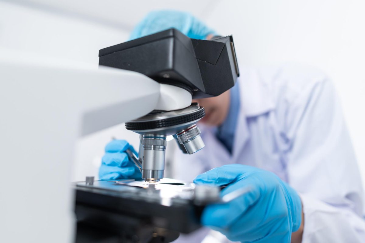
Scientists developed a superlens technique allowing them to see four times smaller than what is capable on a modern microscope. This technique pushed beyond normal microscope limits, breaking the “unbreakable barrier.”
This scientific achievement was seen as nearly impossible because of the diffraction limit, which refers to the physical limit of how closely people can examine objects and organisms.
Objects placed under a modern microscope would have appeared blurry, concealing the object’s size or shape, leaving researchers unable to decipher it. The resolution of microscopes could not capture the light waves at the smallest levels until this discovery.
A team of physicists at the University of Sydney’s press release revealed that their superlensing technique could be used in medical diagnosis and advanced manufacturing with further research.
The improved resolution microscopy can potentially advance imaging techniques in various medical fields such as cancer diagnosis, archaeology, forensics and more.
Alessandro Tuniz, a professor at the School of Physics at USYD, led the research team and discussed how his team measured light at high and low resolutions.
They then entered the operation information on a computer, which selected the most important details while discarding the light waves that would make the image unclear.
The computer program then produced an amplified image of the object’s light waves at four times the original size.
The success of this technique lies in the computer’s ability to select the most vital information while discarding anything that would obscure the image.
Associate professor and researcher on Tuniz’s team, Boris Kuhlmey, found that examining objects from this further distance maintains the “integrity” of the object or, in other words, the accurate size, shape or color.
On the contrary, moving closer to the object or adopting a high-resolution approach would disrupt the image proportions, leading to blurriness.
Their peer-reviewed research paper was published in Nature Communications on Oct. 18, detailing the two main subwavelength imaging techniques used: scattering-type scanning nearfield optical microscopy and near-field photoconductive antennas.
The scanning scatters radiation through an atomic force, leading to the best resolution in today’s age, while the antennas better examine the amplitude, resolving any polarization. Both methods combined allow for the perfect distance, which is close enough to capture the light but not close enough to disrupt the image.
Furthermore, high-resolution optical microscopy will interest people in fields other than in the science sphere. Anything that is hidden by layers can potentially be viewed with this technology.
According to Interesting Engineering, superlens are made from metamaterials, which are connected by different chemical structures and used by scientists for various purposes ranging from filters, devices, applications, detection or monitoring.
Metamaterials are not found in nature and are specifically developed for unique purposes, and in this case, it was developed to push against the diffraction limit.
Any other fields that want to view something blocked by the physical limit of minor light levels would benefit from this new superlens technique.


